
Multimodal MRI processing
T1, FLAIR, diffusion, and functional data can be standardized and aligned for case review.
BrainScores turns routine MRI into quantitative, interpretable maps of structural deviation. Our platform helps physicians identify subtle abnormalities, review evidence at the patient level, and move from imaging insight to treatment planning with confidence.
T1, FLAIR, diffusion, and functional data can be standardized and aligned for case review.

Patient anatomy is compared against reference distributions to show where tissue properties diverge from expected patterns.

Candidate lesions are ranked and visualized so physicians can inspect the underlying anatomy directly.

Outputs can be prepared for neuronavigation and multidisciplinary epilepsy conference review.
Drug-resistant epilepsy often depends on identifying a structural target that conventional reads may miss. BrainScores combines multimodal MRI processing, whole-brain morphometric analysis, AI-assisted lesion detection, and expert validation to surface focal cortical dysplasia, hippocampal abnormalities, atrophy patterns, and other subtle markers relevant to surgical planning.
STANDARD MRI:
Negative
BRAINSCORES ANALYSIS:
Lesion detected in right frontal cortex
SURGICAL OUTCOME:
Seizure-free at 12 months
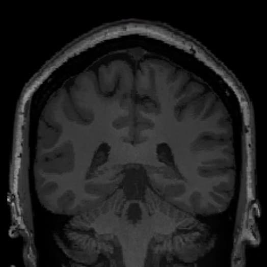
MRI-NEGATIVE
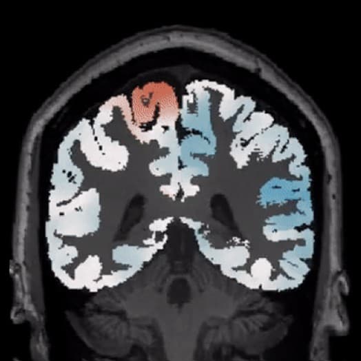
AUTOMATED DEVIATION MAPPING
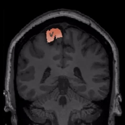
AI DIAGNOSTICS
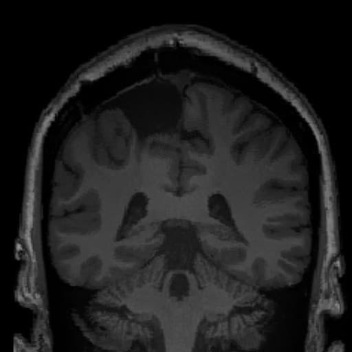
CURATIVE SURGERY
STANDARD MRI:
Negative
BRAINSCORES ANALYSIS:
Hippocampal abnormality identified
SURGICAL OUTCOME:
Seizure-free at 18 months
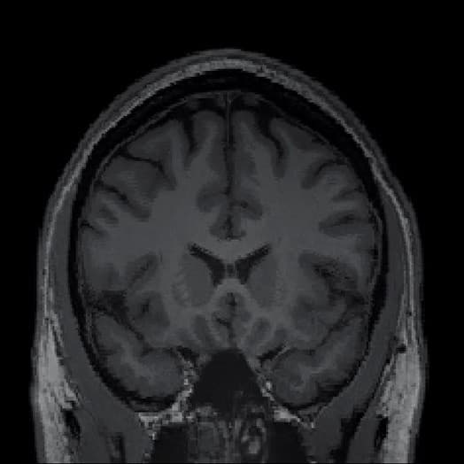
MRI-NEGATIVE
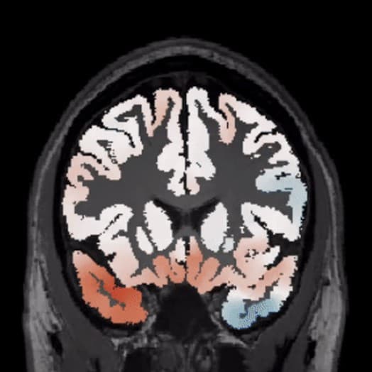
AUTOMATED DEVIATION MAPPING
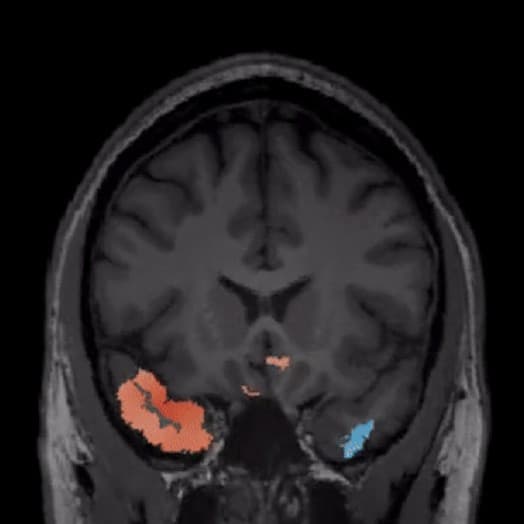
AI DIAGNOSTICS
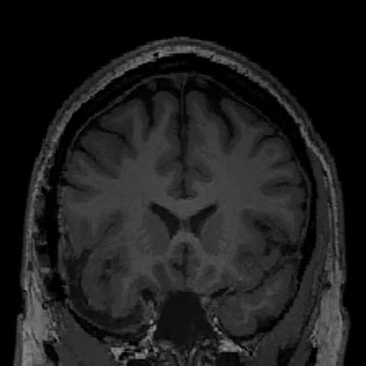
CURATIVE SURGERY
Age-related decline is often gradual, heterogeneous, and difficult to interpret from a single scan. BrainScores can quantify regional brain structure, compare individuals against age-adjusted references, and track patterns that may support earlier recognition, referral, and monitoring.
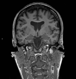
T1 WEIGHTED
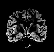
FLAIR
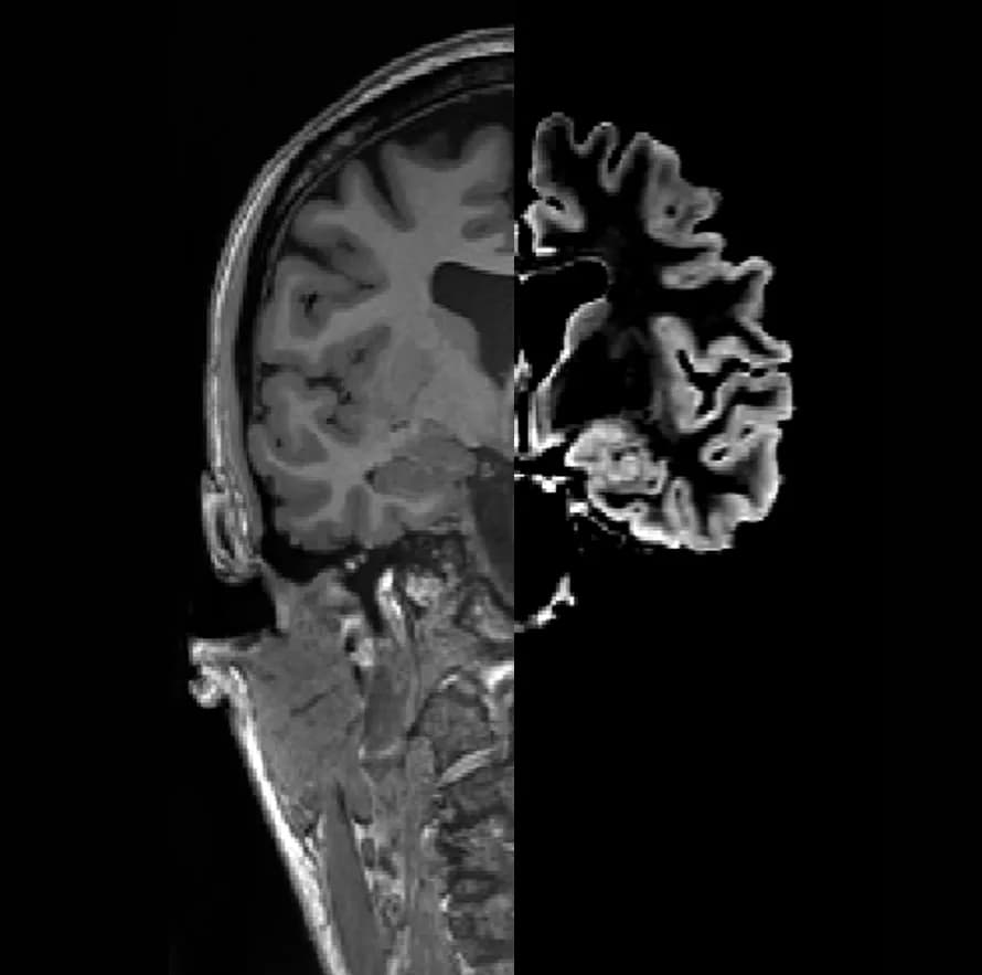
FUSED IMAGES
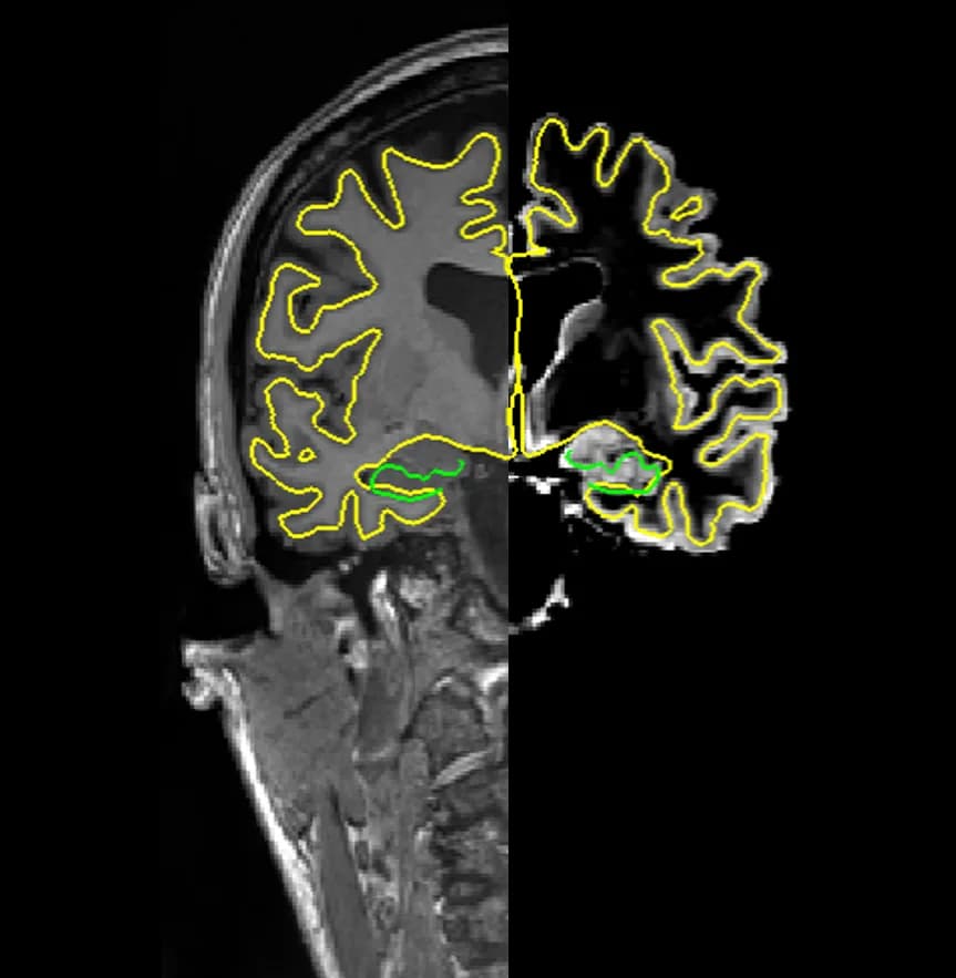
PROCESSED IMAGES
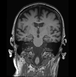
T1 WEIGHTED
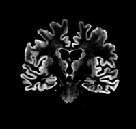
FLAIR
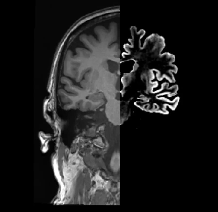
FUSED IMAGES
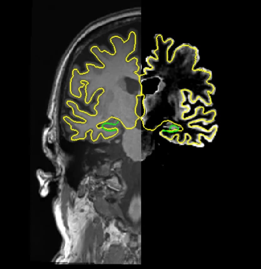
PROCESSED IMAGES
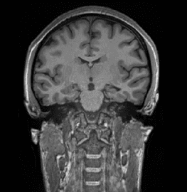
T1 WEIGHTED
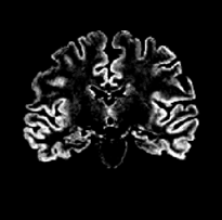
FLAIR
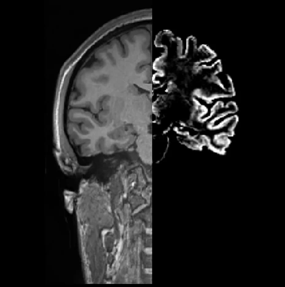
FUSED IMAGES
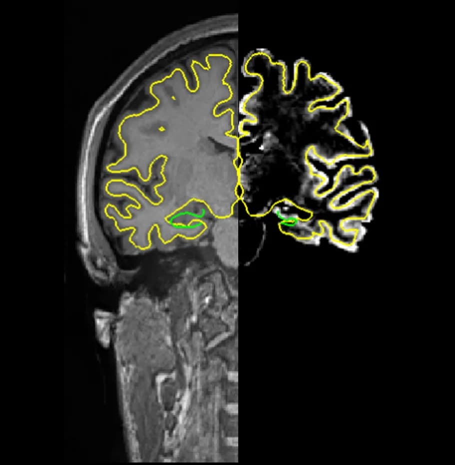
PROCESSED IMAGES
Upload MRI data securely.
BrainScores standardizes imaging and performs automated quantitative mapping.
The platform compares to normative data, produces candidate findings, and visual overlays.
Results can be discussed in clinic, case conference, or surgical planning workflows.
Built by researchers who defined modern epilepsy imaging.
Developed from peer-reviewed algorithms and open-science neuroimaging tools.
Warning
BrainScores outputs are intended to support qualified medical professionals and should be interpreted alongside clinical history, EEG, neuropsychology, radiology, and other standard-of-care evidence.
Talk with our clinical team about MRI-negative epilepsy cases, surgical planning workflows, aging-related imaging questions, or institutional validation studies.